Fat grafting, also known as fat transfers or fat injections, is a plastic surgery procedure which removes fat from unwanted areas of your body, carefully processes the fat, and then reinjects your own adipose fat tissue where more volume is desired. Fat injections can help rejuvenate your face, your buttocks, your hands, or your breasts. Fat injections are also being used in Breast reconstruction surgery. Fat grafting will restore volume to the breast and can be performed with or without an implant.
Lately we have seen some plastic surgery blogs expressing concern regarding how fat injections to the breast affect mammograms. We feel that both sides of the story are not being told, so we would like to address that here.
When we wrote about fat grafting to the breast back in April of 2009, the conclusion from the ASPS position paper indicated no evidence that fat injections interfere with breast cancer detection and that results of fat transfers remain highly dependent on a surgeon’s technique and expertise. See our previous blog post, Fat stem cell injections to the breast – Risky?, for more detail.
Two years later it’s still being debated. The Plastic and Reconstructive Surgery Journal, a journal of peer reviewed studies from the plastic surgery community, recently published several papers that come to different conclusions about whether fat injections to the breast can interfere with cancer screening.
Let’s explore these publications to review the various findings.
The Chinse study

April 2011, Plastic and reconstructive surgery
Clinical analyses of clustered microcalcifications after autologous fat Injection for Breast augmentation
The recently released paper, published in the April 2011 issue, put fat injections to the breast in a negative light. The study was conducted by Chinese plastic surgeons at Meitan General Hospital and Peking Union Medical College Hospital in Beijing, China. The Chinese study concluded that fat injections create issues with mammograms used to detect cancer in the breast. Based on these findings, the study recommended prohibiting all fat injections to the breast.
This study analyzed ‘digitized mammographic films’ from 48 patients who had received fat injections to the breast from July 1999 to December 2009.
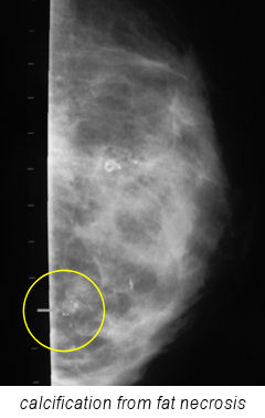
The study focused on calcifications which occur after fat grafting is performed.
When a fat injection does not take well, the fat cells can die (fat necrosis), and take on the appearance of a calcification. The Chinese study concluded that mammograms cannot distinguish between calcifications formed from fat or calcifications that may be cancerous. The study’s abstract conclusion states “clustered microcalcifications found after autologous fat injection for breast augmentation, cannot be distinguished from malignancy.”
Even though all calcifications were biopsied to be non-malignant, the study also reached a grandiose conclusion that fat injections to the breast “should be prohibited.”
So–should fat injections to the breast be outlawed?
Or is there another side to the story? If so, what is it? The heart of the matter is whether radiologists can distinguish if calcifications seen on mammograms are due to dead fat cells, or from malignant cancer cells. First of all, not all fat grafting produces calcifications. If a fat graft takes well, there will be fewer dead fat cells, or fat necrosis, thus resulting in fewer or no calcifications. However, a poorly performed fat graft will likely have lots of dead fat cells, likely resulting in more calcifications.
Let’s look at other recent articles that were published on the topic of fat injections and mammograms, all within the last month.
The discussion article about the Chinese study
April 2011, Plastic and Reconstructive Surgery
Daniel DelVecchio, M.D.
The same April 2011 Plastic Surgery Journal issue included a ‘Discussion’ article about the Chinese study. If you had a hard copy of April’s journal, you would simply turn from the last page of the Chinese publication to read the ‘Discussion’ section that immediately follows written by Plastic Surgeon, Dr. Daniel DelVecchio. Dr. DelVecchio was an early adapter of fat grafting to the breast and has been published frequently in the Plastic Surgery literature.
Dr. DelVecchio brought up several excellent points as to why the Chinese publication conclusion was without merit. One point addressed concerned the issue of the resolution quality of the mammograms. What type of mammography did the Chinese use? The Chinese publications stated that they used ‘digitized mammographic films.’
screen film mammograms are inferior to direct digital mammograms
According to DelVecchio, this would imply that plain screen film mammograms were initially imaged, and later digitized. Per DelVecchio: “the quality of digitized screen film mammogram is only as good as the quality of the original mammogram, and it is common industry knowledge that screen film mammograms are inferior to direct digital mammograms.”
In other words – did the Chinese study simply digitize mammogram films with inferior resolution?
Dr. DelVecchio pointed to the work of other fat grafting pioneers, such as Dr. Emmanuel Delay, who have described the ability to distinguish between benign and malignant findings on post fat grafting mammograms. DelVecchio cited the radiologic literature which supported the general concept that calcifications and fat necrosis can be distinguished from the malignant signs of irregularly shaped, high-opacity microcalcifications when supplemental imaging modalities are considered.
Other notable points made by Dr. DelVecchio included the need for standardization of fat injection procedures in order to minimize fat necrosis, which result in calcifications. Deviation from ideal fat grafting techniques result in poor fat take because some of the fat dies. It is these dead fat cells that can result in calcifications. As standards emerge for the best fat grafting techniques, there will be less fat necrosis and calcifications to show up on mammograms as a result of fat injections to the breast.
In DelVecchio’s opinion, modern mammography techniques can distinguish between malignancy and benign fat deposits found on mammograms.
The French study

March 2011, Plastic and Reconstructive Surgery
Radiographic findings after Breast augmentation by autologous fat transfer
Exactly one month prior to the Chinese article publication, a group of French surgeons published an article in the March 2011 issue of Plastic Surgery Journal. The study compared mammograms pre and post operative mammograms on 20 patients who underwent fat grafting to the breast. The study’s goal was to determine whether the transfer of fat to the native breast hampers breast imaging.
Using guidelines stabled by the American College of Radiology classification, the study concluded that radiographic follow up of breast treated with fat grafting is not problematic and should not be a hindrance to the procedures. However, the study did recommend that techniques used for fat injection patients should become standardized, so that reproducibility could be insured.
Our summary
As more radiographers gain experience with fat grafting, we expect this mammogram issue to diminish, as happened with the case of breast implants and mammography.
Additionally, there is one important fact to remember–of all the thousands of fat grafts to the breast that have been done to present, there is yet to be a reported case of a cancer being missed on a mammogram because of a previous fat graft.


buttock injections says:
Alex says:
Dr. Ricardo L Rodriguez says:
Deucey says:
Dr. Ricardo L Rodriguez says:
Deb says:
Dr. Ricardo L Rodriguez says:
Sussie says:
Dr. Ricardo L Rodriguez says:
Joseph Stevens says:
Dr. Ricardo L Rodriguez says:
Dean Greg says:
Dr. Ricardo L Rodriguez says: